Prophylactic Antibiotic Use in Adult Cardiac Arrest Patients Receiving Emergent ECMO (eCPR)
Introduction
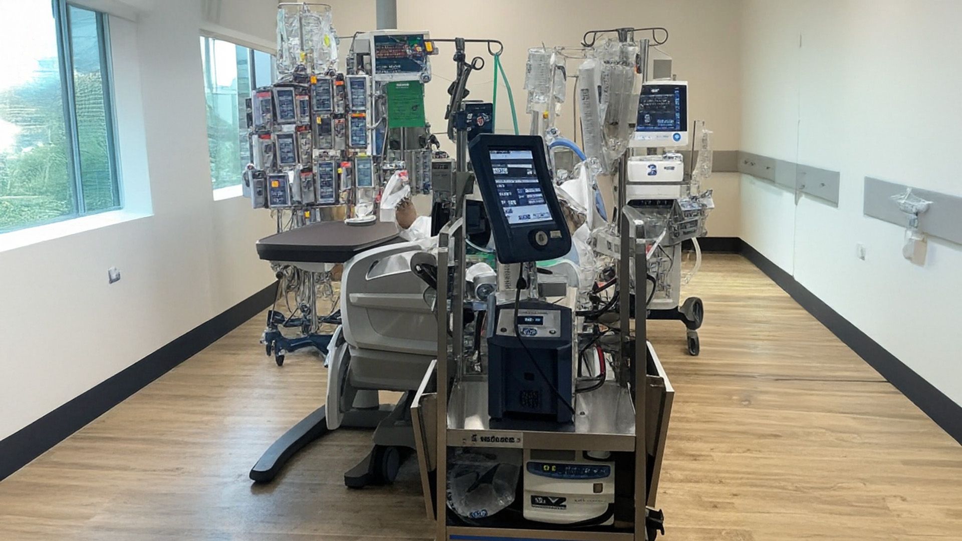
Extracorporeal Membrane Oxygenation (ECMO) is an advanced life support therapy utilized in critical care medicine to provide temporary circulatory and respiratory support for patients with severely compromised cardiac and/or pulmonary function.1 In the context of cardiac arrest, the rapid deployment of ECMO, termed extracorporeal cardiopulmonary resuscitation (eCPR), aims to restore systemic perfusion and improve survival rates in patients who do not respond to conventional resuscitation efforts.2 While ECMO offers a crucial bridge to recovery, it inherently carries a significant risk of infection. The establishment of the ECMO circuit necessitates the insertion of large-bore cannulas into major blood vessels, a procedure that, particularly in the emergent setting of cardiac arrest, may be conducted under less than ideal sterile conditions, thereby increasing the potential for microbial contamination at the insertion site. Furthermore, the ECMO circuit itself, involving invasive cannulas that breach the skin’s natural protective barrier, provides a direct pathway for pathogenic microorganisms to enter the bloodstream.4 The complexity of care for patients on ECMO often involves multiple additional invasive interventions, such as central venous catheters for medication administration and arterial lines for hemodynamic monitoring, which further elevate the susceptibility to hospital-acquired infections.4 This report aims to critically evaluate the current recommendations and available evidence regarding the use of prophylactic antibiotics in adult patients who experience cardiac arrest and are emergently placed on ECMO. It will specifically address the concerns arising from the potentially semi-sterile nature of cannula insertion during eCPR and the implications of prolonged ECMO support on infection risk.
The urgent need to establish circulatory support during eCPR might sometimes necessitate a more rapid cannulation procedure, potentially with less stringent adherence to sterility protocols compared to elective ECMO initiation. This reality could contribute to a higher initial risk of infections at the cannula insertion sites. Additionally, patients who survive the initial cardiac arrest and are supported by ECMO often require prolonged treatment, sometimes lasting for days or even weeks.1 This extended duration of ECMO support, with indwelling cannulas and other invasive devices, progressively increases the cumulative risk of various nosocomial infections, extending beyond just the initial insertion site.
Current Guidelines on Prophylactic Antibiotics in ECMO
The Extracorporeal Life Support Organization (ELSO) provides guidelines for the management of patients receiving ECMO. Current ELSO recommendations generally advise against the routine use of antimicrobial prophylaxis in ECMO patients to prevent infections, beyond the standard periprocedural antibiotic administration.7 This stance is primarily based on the observation that retrospective studies investigating the efficacy of routine prophylactic antibiotics after ECMO cannulation have not consistently demonstrated a clear benefit in improving patient outcomes.9 ELSO guidance suggests the administration of only standard periprocedural antimicrobial prophylaxis, which is typically a single dose given shortly before the procedure to prevent surgical site infections from skin commensals.9
While ELSO offers various guidelines covering different aspects of ECMO support, including specific guidelines for adult and pediatric eCPR 10, the provided research material does not detail a specific recommendation within these guidelines regarding routine antibiotic prophylaxis in the eCPR setting. It is noted that ELSO has developed in-depth guidelines for ECMO in COVID-19 patients 10, which likely include detailed infection prevention and control strategies tailored to that specific patient population. However, the applicability of these COVID-19-specific recommendations to the broader population of non-COVID eCPR patients requires careful consideration of the differing risk factors and clinical contexts.
The rationale behind ELSO’s discouragement of routine antibiotic prophylaxis in ECMO patients stems from the inconsistent findings of retrospective studies, which have failed to demonstrate a consistent improvement in patient outcomes with this practice.9 Furthermore, there are significant concerns about the potential for routine antibiotic use to promote the development of antibiotic resistance, a critical issue in the management of critically ill patients who are already at high risk of infection with multidrug-resistant organisms.11
The lack of a specific ELSO recommendation either endorsing or opposing routine prophylactic antibiotics in eCPR suggests that the general guidance for ECMO patients should be applied. However, the unique circumstances and potential risks associated with eCPR, such as the possibility of less than ideal sterile conditions during emergent cannulation, might necessitate a more nuanced approach at the individual institutional level. Each center should consider its local infection rates, prevalent pathogens, and patient risk factors when developing protocols for infection prevention in eCPR patients.
Evidence for Prophylactic Antibiotics in eCPR Patients
The evidence regarding the effectiveness of prophylactic antibiotics in ECMO patients, including those undergoing eCPR, is varied and often conflicting. A nationwide cohort study conducted in Japan by Kondo et al. 14 analyzed a large database of patients receiving ECMO and found that the administration of prophylactic antibiotics (specifically cephalosporins or glycopeptides) within the first two days of ECMO initiation was associated with a reduction in in-hospital mortality and a lower incidence of nosocomial pneumonia. However, this study had several limitations, including the fact that it did not specifically focus on patients who underwent eCPR and the definition of prophylaxis included antibiotic administration up to 48 hours after ECMO initiation, which might blur the line between true prophylaxis and early empirical treatment.13 Additionally, the study did not provide detailed information on the specific antibiotic agents used, their dosages, or the duration of treatment.
In contrast, a systematic review published in 2016, which evaluated 11 studies on antibiotic prophylaxis in ECMO patients, concluded that infection rates were generally similar regardless of prophylaxis use, and the two studies that directly compared outcomes with and without prophylaxis found no benefit from routine antibiotic administration.7 The review also noted the heterogeneity of causative infectious organisms, which did not support a clear rationale for any specific prophylactic strategy.
However, a more recent retrospective study specifically focused on patients undergoing eCPR found a potential benefit of prophylactic antibiotics with coverage against Pseudomonas aeruginosa.3 This study observed that patients who received prophylactic antibiotics with Pseudomonas coverage within 72 hours after eCPR had a significantly lower incidence of nosocomial infections, particularly ventilator-associated pneumonia (VAP), where Pseudomonas was a common pathogen. The antibiotics with Pseudomonas coverage used in this study included ceftazidime, cefepime, ciprofloxacin, levofloxacin, and meropenem.20 This finding suggests that in the eCPR population, where VAP is a frequent complication and Pseudomonas is often implicated, targeted prophylaxis might be beneficial.
A systematic review from 2024, encompassing three studies with a large number of ECMO participants, indicated that prophylactic antibiotics were associated with a reduction in mortality, the risk of infections, and complications such as acute kidney injury and diarrhea.18 However, the authors of this review also emphasized the need for further prospective research and the development of tailored, ECMO-specific bundles to optimize infection prevention strategies.
The available evidence presents a complex picture. While some studies, particularly the large Japanese cohort and the eCPR-specific study on Pseudomonas coverage, suggest potential benefits of prophylactic antibiotics in certain contexts, the overall evidence is not robust enough to support a universal recommendation for routine use in all adult patients receiving eCPR. The heterogeneity in study designs, patient populations, antibiotic regimens, and outcome measures contributes to the conflicting findings.
Table 1: Summary of Key Studies Investigating Prophylactic Antibiotics in ECMO
| Study Author and Year | Study Design | Patient Population | Intervention | Comparison Group | Main Findings |
| Kondo et al., 2021 | Retrospective Cohort | Adults (general ECMO) | Cephalosporin or glycopeptide within 2 days of ECMO | No antibiotics | Reduced in-hospital mortality and nosocomial pneumonia associated with prophylaxis. |
| Systematic Review, 2016 | Systematic Review | Adults, Children, Neonates (general ECMO) | Various prophylactic antibiotic regimens | No prophylaxis | No benefit from routine antibiotic prophylaxis in most patients; heterogeneous causative organisms. |
| PMC10774957, 2023 | Retrospective Cohort | Adults (eCPR) | Prophylactic antibiotics with Pseudomonas coverage | No prophylaxis | Lower incidence of nosocomial infections, particularly VAP, associated with Pseudomonas coverage within 72 hours of eCPR. |
| Systematic Review, 2024 | Systematic Review | Adults (general ECMO) | Various prophylactic antibiotic regimens | No prophylaxis | Prophylactic antibiotics associated with reduced mortality, infections, and complications, but calls for more prospective research. |
IV. Rationale Against Routine Prophylaxis and Considerations for Antimicrobial Stewardship
The routine use of prophylactic antibiotics in ECMO patients carries significant potential risks. One of the most concerning is the increased likelihood of developing antibiotic resistance among both the patient’s own flora and the pathogens responsible for subsequent infections.1 This can lead to infections that are more difficult to treat and may require the use of broader-spectrum antibiotics, further exacerbating the problem of resistance. Additionally, antibiotic prophylaxis can disrupt the normal microbial balance in the body, potentially leading to opportunistic infections such as Clostridioides difficile infection.11 Patients may also experience direct drug toxicities or other adverse side effects from the prophylactic antibiotics themselves.12 Furthermore, the fear of missing an infection in this complex patient population can sometimes lead to the overdiagnosis of ECMO-associated infections (EAIs), resulting in the prolonged use of broad-spectrum antibiotics, which further contributes to the risks of resistance and C. difficile infection.11
Given these potential harms and the limited evidence of clear benefit for routine prophylaxis in ECMO, the principles of antimicrobial stewardship are of paramount importance in this vulnerable patient population.1 Antimicrobial stewardship aims to optimize antibiotic use to improve patient outcomes while minimizing the development of resistance. In the context of eCPR, this means avoiding unnecessary antibiotic exposure unless there is strong evidence to support its use in a specific situation.1 If prophylactic antibiotics are considered, the choice of agent should be carefully tailored based on the local microbiology and antibiotic resistance patterns prevalent in the institution.11 It is also crucial to regularly review and revise antibiotic protocols based on local infection data and emerging resistance trends.11
The potential for routine antibiotic prophylaxis to promote antibiotic resistance likely outweighs the uncertain benefits, especially in the absence of robust evidence. This highlights the critical need for a judicious approach to antibiotic use in ECMO patients, prioritizing antimicrobial stewardship to protect both individual patients and the broader healthcare system from the consequences of antibiotic resistance.
Potential Role of Targeted Prophylaxis in Specific Subgroups or Settings
While ELSO generally advises against routine antibiotic prophylaxis in ECMO patients, there might be specific patient characteristics or procedural factors that could warrant consideration for a more targeted approach in the eCPR setting. Immunocompromised patients, for instance, may be at a higher baseline risk of infection.1 ELSO guidelines acknowledge that prophylaxis might be considered in immunocompromised individuals.13 However, the specific definition of immunocompromised in this context and the optimal prophylactic regimen remain unclear and require careful clinical judgment.
ELSO also mentions patients with a prolonged open chest, often encountered after cardiac surgery, as a potential indication for prophylactic antibiotics.7 This scenario is less directly applicable to eCPR unless the cannulation was performed via an open chest approach, which is less common than percutaneous insertion.
Regarding procedural factors, central cannulation has been suggested to potentially carry a higher risk of certain infections compared to peripheral cannulation.1 However, current guidelines do not differentiate prophylaxis recommendations based on the site of cannulation, and further evidence is needed to determine if targeted prophylaxis is warranted in patients undergoing central cannulation for eCPR.
Although emergent cannulation during eCPR might theoretically be associated with a higher risk of infection due to potentially less controlled sterile conditions, current guidelines do not distinguish prophylaxis recommendations based on the urgency of the procedure. The decision to consider targeted prophylaxis in specific subgroups of eCPR patients should be based on a comprehensive assessment of the individual patient’s risk factors, the specific circumstances of their cardiac arrest and resuscitation, and the local epidemiology of infections in the ECMO unit.
Importance of Infection Prevention and Control Measures
Comprehensive infection prevention and control measures are paramount in reducing the risk of nosocomial infections in ECMO patients and are likely more impactful than routine antibiotic prophylaxis. Meticulous adherence to aseptic techniques during ECMO cannulation and the ongoing care of cannula insertion sites are fundamental.1 Implementing standardized cannula site care bundles, adapted from principles used for central line-associated bloodstream infection (CLABSI) prevention, has shown significant success in reducing infection rates.5 These bundles typically focus on standardizing the use of antimicrobial dressings, ensuring strict aseptic techniques during dressing changes and line access, optimizing cannula securement to prevent movement and disruption of the insertion site, performing daily antimicrobial patient bathing, and addressing environmental factors to minimize contamination risks.31
Routine monitoring of cannula insertion sites for any signs of infection, such as erythema, purulent drainage, or localized tenderness, is essential.1 Maintaining a low threshold to obtain cultures from blood and insertion sites when infection is suspected allows for early identification of pathogens and timely initiation of appropriate treatment.1 Efforts should also be made to avoid maintaining ECMO treatment for longer than absolutely necessary, as prolonged duration of support is a well-established risk factor for infection.1 Other important infection control practices include the use of needleless hubs at all connections in the ECMO circuit to reduce the risk of contamination 4, the routine disinfection of ECMO system parts with chlorhexidine 4, and ensuring frequent hand hygiene by all healthcare personnel involved in the care of the patient.4
The experience of centers that have implemented comprehensive infection prevention bundles has demonstrated remarkable success in significantly reducing bloodstream infections in ECMO patients.31 These success stories underscore the effectiveness of a multi-faceted approach that focuses on meticulous care practices and standardized protocols to minimize the risk of infection.
Considerations for Antibiotic Regimens if Prophylaxis is Used
If, despite the general lack of strong evidence, a clinician decides to use prophylactic antibiotics in an adult patient receiving eCPR, the choice of antibiotic regimen should be carefully considered. The Japanese cohort study mentioned the use of cephalosporins or glycopeptides within the first two days of ECMO initiation.14 However, the specific agents, dosages, and durations were not detailed in the provided snippets. The eCPR study suggesting a benefit of Pseudomonas coverage did not specify a particular regimen but mentioned classes of antibiotics that have activity against Pseudomonas, including ceftazidime, cefepime, ciprofloxacin, levofloxacin, and meropenem.20 It is important to note that any consideration of these agents for prophylaxis should be guided by the local antibiogram, which provides information on the susceptibility patterns of common pathogens in a specific institution. Patient-specific factors, such as allergies and prior antibiotic exposure, should also be taken into account. One study 25 reported on the development of a standardized antimicrobial prophylaxis regimen for ECMO patients based on their institutional experience, but the specific details of this regimen are not available in the provided material.
It is crucial to emphasize that there is no universally recommended antibiotic regimen for prophylactic use in eCPR patients, and any decision to use prophylaxis should be made on an individualized basis, considering the potential benefits and risks in the context of the specific clinical situation and local microbiological data. Furthermore, it is important to acknowledge the significant challenges associated with antibiotic dosing in ECMO patients due to the pharmacokinetic and pharmacodynamic alterations that can occur with ECMO support.30 Factors such as increased volume of distribution, altered drug clearance, and potential sequestration of antibiotics within the ECMO circuit can affect drug levels and efficacy. Therefore, if prophylactic antibiotics are used, ensuring therapeutic levels is important, and in cases of suspected or confirmed infection, therapeutic drug monitoring may be warranted.
Table 2: Antibiotics with Pseudomonas aeruginosa Coverage Mentioned in eCPR Study
| Antibiotic Name | Class of Antibiotic |
| Ceftazidime | Cephalosporin |
| Cefepime | Cephalosporin |
| Ciprofloxacin | Fluoroquinolone |
| Levofloxacin | Fluoroquinolone |
| Meropenem | Carbapenem |
VIII. Conclusion and Recommendations
In summary, the current evidence regarding the use of prophylactic antibiotics in adult patients receiving emergent ECMO for cardiac arrest (eCPR) is not robust enough to support a routine recommendation. While a large retrospective study suggested a potential benefit of early antibiotic administration in ECMO patients in general, and a study focused on eCPR indicated that prophylactic antibiotics with Pseudomonas aeruginosa coverage might reduce nosocomial infections, the overall evidence remains limited and often conflicting. The Extracorporeal Life Support Organization (ELSO) generally recommends against routine antimicrobial prophylaxis in ECMO patients, citing the lack of clear patient outcome benefit and concerns about promoting antibiotic resistance.
Given the absence of strong evidence supporting routine prophylaxis and the significant risks associated with widespread antibiotic use, a nuanced approach is warranted. While routine prophylactic antibiotics are not recommended for all adult patients receiving eCPR, there might be specific contexts or patient subgroups where targeted prophylaxis could be considered. For instance, based on the findings of a study focused on eCPR, prophylactic antibiotics with coverage against Pseudomonas aeruginosa within the first 72 hours of ECMO initiation might be considered, especially in institutions with a high prevalence of Pseudomonas VAP. However, this decision should be made with caution and in the context of local antibiograms and a thorough assessment of the individual patient’s risk factors. Patients with known immunocompromising conditions might also warrant consideration for targeted prophylaxis, although specific regimens and durations require further investigation.
The cornerstone of reducing ECMO-associated infections in eCPR patients should be the meticulous implementation and consistent adherence to comprehensive infection prevention bundles. These bundles, adapted from best practices for CLABSI prevention, should focus on standardized protocols for cannula insertion and maintenance, including aseptic techniques, antimicrobial dressings, cannula securement, and routine site monitoring. Furthermore, strict adherence to antimicrobial stewardship principles is crucial to minimize unnecessary antibiotic use and mitigate the development of antibiotic resistance.
Future research should focus on conducting prospective, well-designed studies to evaluate the efficacy of targeted prophylactic strategies in specific subgroups of eCPR patients. Identifying patient and procedural factors that are associated with a higher risk of infection could help to refine prophylactic approaches and optimize infection prevention strategies in this complex and critically ill population.
Reference
- Infection Control Challenges and Extracorporeal Membrane Oxygenation – UTMB Health, accessed April 26, 2025, https://www.utmb.edu/spectre/news-events/all-news/article/spectre-blog/2023/12/04/infection-control-challenges-and-extracorporeal-membrane-oxygenation
- What Is ECMO? | Extracorporeal Membrane Oxygenation (ECMO), accessed April 26, 2025, https://elso.org/extracorporeal-membrane-oxygenation.aspx
- (PDF) Prophylactic antibiotic treatment for preventing nosocomial infection in extracorporeal membrane oxygenation-resuscitated circulatory arrest patients – ResearchGate, accessed April 26, 2025, https://www.researchgate.net/publication/373340228_Prophylactic_antibiotic_treatment_for_preventing_nosocomial_infection_in_extracorporeal_membrane_oxygenation-resuscitated_circulatory_arrest_patients
- Prevention of catheter-related bloodstream infections in patients with extracorporeal membrane oxygenation: a literature review – PubMed Central, accessed April 26, 2025, https://pmc.ncbi.nlm.nih.gov/articles/PMC10511280/
- Prevention of catheter-related bloodstream infections in patients with extracorporeal membrane oxygenation: a literature review – SciELO, accessed April 26, 2025, https://www.scielo.br/j/ramb/a/TSwSJvR9HKqCjrpFgD8LKrh/?lang=en
- Prevention of catheter-related bloodstream infections in patients with extracorporeal membrane oxygenation: a literature review – SciELO, accessed April 26, 2025, https://www.scielo.br/j/ramb/a/TSwSJvR9HKqCjrpFgD8LKrh/?format=pdf&lang=en
- (PDF) The Evidence Base for Prophylactic Antibiotics in Patients Receiving Extracorporeal Membrane Oxygenation – ResearchGate, accessed April 26, 2025, https://www.researchgate.net/publication/282874009_The_Evidence_Base_for_Prophylactic_Antibiotics_in_Patients_Receiving_Extracorporeal_Membrane_Oxygenation
- The Evidence Base for Prophylactic Antibiotics in Patients Receiving Extracorporeal Membrane Oxygenation – ResearchGate, accessed April 26, 2025, https://www.researchgate.net/profile/Alice-Gallo-De-Moraes/publication/282874009_The_Evidence_Base_for_Prophylactic_Antibiotics_in_Patients_Receiving_Extracorporeal_Membrane_Oxygenation/links/5a5fc442aca27273524581ec/The-Evidence-Base-for-Prophylactic-Antibiotics-in-Patients-Receiving-Extracorporeal-Membrane-Oxygenation.pdf
- Nosocomial Infections in Adults Receiving Extracorporeal Membrane Oxygenation: A Review for Infectious Diseases Clinicians – Oxford Academic, accessed April 26, 2025, https://academic.oup.com/cid/article/79/2/412/7621752
- ELSO Guidelines – Extracorporeal Life Support Organization, accessed April 26, 2025, https://www.elso.org/ecmo-resources/elso-ecmo-guidelines.aspx
- Infectious Diseases Provider Role in Extracorporeal Membrane Oxygenation Infections, accessed April 26, 2025, https://www.contagionlive.com/view/infectious-diseases-provider-role-in-extracorporeal-membrane-oxygenation-infections
- News Scan for Feb 12, 2021 – CIDRAP, accessed April 26, 2025, https://www.cidrap.umn.edu/news-scan-feb-12-2021
- Antimicrobial Prophylaxis in Extracorporeal Membrane Oxygenation: Is the Debate Still Open? – PMC, accessed April 26, 2025, https://pmc.ncbi.nlm.nih.gov/articles/PMC9116348/
- Efficacy of Prophylactic Antibiotics during Extracorporeal Membrane Oxygenation: A Nationwide Cohort Study | Annals of the American Thoracic Society, accessed April 26, 2025, https://www.atsjournals.org/doi/10.1513/AnnalsATS.202008-974OC
- Efficacy of Prophylactic Antibiotics during Extracorporeal Membrane Oxygenation: A Nationwide Cohort Study | Annals of the American Thoracic Society, accessed April 26, 2025, https://www.atsjournals.org/doi/full/10.1513/AnnalsATS.202008-974OC
- Efficacy of Prophylactic Antibiotics during Extracorporeal Membrane Oxygenation: A Nationwide Cohort Study – PubMed, accessed April 26, 2025, https://pubmed.ncbi.nlm.nih.gov/33765406/
- Antimicrobial Prophylaxis in Extracorporeal Membrane Oxygenation: Is the Debate Still Open? | Annals of the American Thoracic Society, accessed April 26, 2025, https://www.atsjournals.org/doi/full/10.1513/AnnalsATS.202201-042LE
- (PDF) Antibiotic Prophylaxis in Patients On Extracorporeal Membrane Oxygenation: A Systematic Review – ResearchGate, accessed April 26, 2025, https://www.researchgate.net/publication/379081197_Antibiotic_Prophylaxis_in_Patients_On_Extracorporeal_Membrane_Oxygenation_A_Systematic_Review
- The Evidence Base for Prophylactic Antibiotics in Patients Receiving Extracorporeal Membrane Oxygenation – PubMed, accessed April 26, 2025, https://pubmed.ncbi.nlm.nih.gov/26461238/
- Prophylactic antibiotic treatment for preventing nosocomial infection …, accessed April 26, 2025, https://pmc.ncbi.nlm.nih.gov/articles/PMC10774957/
- Prophylactic antibiotic treatment for preventing nosocomial infection in extracorporeal membrane oxygenation-resuscitated circulatory arrest patients – PubMed, accessed April 26, 2025, https://pubmed.ncbi.nlm.nih.gov/38204699/
- Antibiotic Prophylaxis in Patients On Extracorporeal Membrane Oxygenation: A Systematic Review – AmSECT, accessed April 26, 2025, https://amsect.org/events-education/calendar-of-events/event-details/antibiotic-prophylaxis-in-patients-on-extracorporeal-membrane-oxygenation-a-systematic-review
- Antibiotic Prophylaxis in Patients on Extracorporeal Membrane Oxygenation: A Systematic Review – PubMed, accessed April 26, 2025, https://pubmed.ncbi.nlm.nih.gov/38502730/
- Infection in ECMO patients: Changes in epidemiology, diagnosis and prevention – PubMed, accessed April 26, 2025, https://pubmed.ncbi.nlm.nih.gov/37925153/
- Reducing Broad-Spectrum Antimicrobial Use in Extracorporeal Membrane Oxygenation: Reduce AMMO Study | Clinical Infectious Diseases | Oxford Academic, accessed April 26, 2025, https://academic.oup.com/cid/article-abstract/73/4/e988/6133715
- Q&A: Best Practices for Infection Prevention in ECMO – Physician’s Weekly, accessed April 26, 2025, https://www.physiciansweekly.com/qa-best-practices-for-infection-prevention-in-ecmo/
- Risk factors for nosocomial infection in patients undergoing extracorporeal membrane oxygenation support treatment: A systematic review and meta-analysis – PubMed, accessed April 26, 2025, https://pubmed.ncbi.nlm.nih.gov/39585868/
- Risk factors for nosocomial infection in patients undergoing extracorporeal membrane oxygenation support treatment: A systematic review and meta-analysis | PLOS One, accessed April 26, 2025, https://journals.plos.org/plosone/article?id=10.1371/journal.pone.0308078
- Antimicrobial Prophylaxis in Extracorporeal Membrane Oxygenation: Is the Debate Still Open? – ATS Journals, accessed April 26, 2025, https://www.atsjournals.org/doi/pdf/10.1513/AnnalsATS.202111-1290LE?download=true
- Antimicrobials – Alfred ECMO Guideline, accessed April 26, 2025, https://ecmo.icu/daily-care-organ-support-in-ecmo-antimicrobials/
- To Eliminate COVID-Era Bloodstream Infections in Heart, Lung Patients, Tampa Hospital Had to Innovate – APIC, accessed April 26, 2025, https://apic.org/to-eliminate-covid-era-bloodstream-infections-in-heart-lung-patients-tampa-hospital-had-to-innovate/
- A Bundled Approach to Integrative Care for Peripherally Inserted Extracorporeal Membrane Oxygenation Cannula Insertion Site – OpenRiver, accessed April 26, 2025, https://openriver.winona.edu/cgi/viewcontent.cgi?article=1040&context=nursingdnp
- Tailored ECMO care bundle eliminated bloodstream infections at Florida hospital – Healio, accessed April 26, 2025, https://www.healio.com/news/infectious-disease/20240603/tailored-ecmo-care-bundle-eliminated-bloodstream-infections-at-florida-hospital
- A Bundled Approach to Integrative Care for Peripherally Inserted Extracorporeal Membrane Oxygenation Cannula Insertion Site – OpenRiver, accessed April 26, 2025, https://openriver.winona.edu/nursingdnp/41/
- Bundle approach used to achieve zero central line-associated bloodstream infections in an adult coronary intensive care unit, accessed April 26, 2025, https://pmc.ncbi.nlm.nih.gov/articles/PMC7893645/
- CLABSI Bundle Results – AACN, accessed April 26, 2025, https://www.aacn.org/newsroom/clabsi-bundle-results
- Bloodstream infection event (central line-associated bloodstream infection and non-central line–associated bloodstream infection) – CDC, accessed April 26, 2025, https://www.cdc.gov/nhsn/pdfs/pscmanual/4psc_clabscurrent.pdf
- Analysis of Nosocomial Infection and Risk Factors in Patients with ECMO Treatment – PMC, accessed April 26, 2025, https://pmc.ncbi.nlm.nih.gov/articles/PMC8241808/
- Outcome and Clinical Characteristics of Nosocomial Infection in Adult Patients Undergoing Extracorporeal Membrane Oxygenation: A Systematic Review and Meta-Analysis – Frontiers, accessed April 26, 2025, https://www.frontiersin.org/journals/public-health/articles/10.3389/fpubh.2022.857873/full
- Extracorporeal Membrane Oxygenation-Related Nosocomial Infection after Cardiac Surgery in Adult Patients – SciELO, accessed April 26, 2025, https://www.scielo.br/j/rbccv/a/9xDSH7LX3qz8q6F6bLwxL4b/?lang=en&format=pdf
- Nosocomial Infections During Extracorporeal Membrane Oxygenation in Pediatric Patients: A Multicenter Retrospective Study – Frontiers, accessed April 26, 2025, https://www.frontiersin.org/journals/pediatrics/articles/10.3389/fped.2022.873577/full
- Incidence, risk factors and outcomes of nosocomial infection in adult patients supported by extracorporeal membrane oxygenation: a systematic review and meta-analysis, accessed April 26, 2025, https://pmc.ncbi.nlm.nih.gov/articles/PMC11088079/
- Epidemiology and risk factors of infections among patients with extracorporeal membrane oxygenation in a tertiary heart center, accessed April 26, 2025, https://www.europeanreview.org/wp/wp-content/uploads/7235-7244.pdf
- Epidemiology and risk factors of infections among patients with extracorporeal membrane oxygenation in a tertiary heart center – European Review for Medical and Pharmacological Sciences, accessed April 26, 2025, https://www.europeanreview.org/article/33295
- Full article: Analysis of Nosocomial Infection and Risk Factors in Patients with ECMO Treatment – Taylor & Francis Online, accessed April 26, 2025, https://www.tandfonline.com/doi/full/10.2147/IDR.S306209
- Nosocomial Infections on ECMO: Unraveling Risk Factors and Impact on Patient Outcomes, accessed April 26, 2025, https://www.physiciansweekly.com/nosocomial-infections-on-ecmo-unraveling-risk-factors-and-impact-on-patient-outcomes/
- Tampa General Hospital’s Success in Eliminating Bloodstream Infections in ECMO Patients, accessed April 26, 2025, https://www.infectioncontroltoday.com/view/tampa-general-hospital-s-success-eliminating-bloodstream-infections-in-ecmo-patients
- Northwestern Medicine Antimicrobial ECMO Dosing Guidance, accessed April 26, 2025, https://adsp.nm.org/uploads/1/4/3/0/143064172/nm_antimicrobial_ecmo_dosing.pdf
- Antibiotics and ECMO in the Adult Population—Persistent Challenges and Practical Guides, accessed April 26, 2025, https://pmc.ncbi.nlm.nih.gov/articles/PMC8944696/
- Antibiotics and ECMO in the Adult Population—Persistent Challenges and Practical Guides, accessed April 26, 2025, https://www.mdpi.com/2079-6382/11/3/338


