
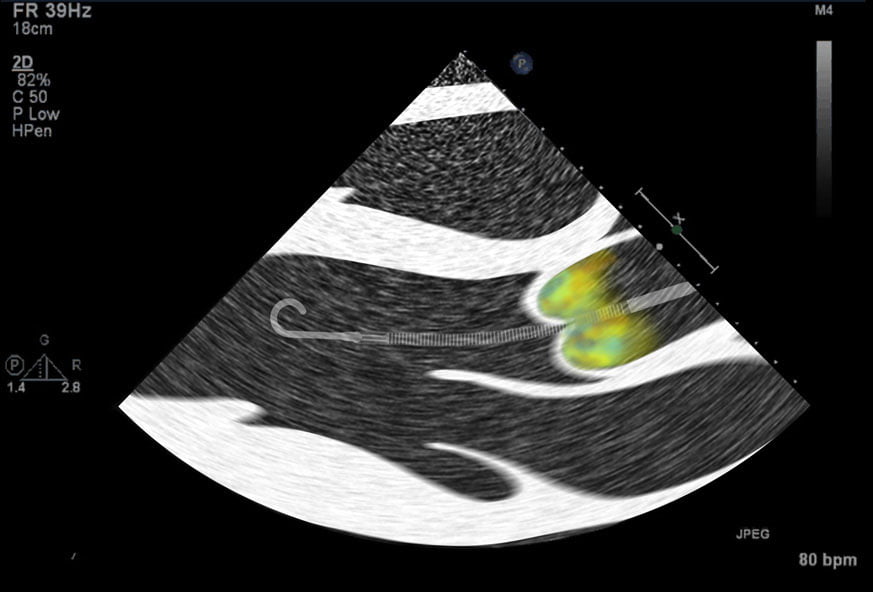
Impella Catheter Positioning
Four Catheter positions you are likely to encounter when examining echocardiograms from patients supported with the Impella CP Catheter.
Calculates stroke volume, cardiac output and cardiac index from the left ventricular outflow tract (LVOT) diameter and velocity-time integral (VTI) on transthoracic echo.
Body size, for indexed values
| Value | Reference | Source |
|---|---|---|
| Cardiac index | 2.5-4.0 L/min/m2 | Normal at right heart catheterization (ESC/ERS 2022) |
| Stroke volume | 60-100 mL | Same |
| Stroke volume index | 33-47 mL/m2 | Same |
| LVOT VTI | Below 16 cm | The most important echo cut-off for hospital mortality in a decision-tree analysis of 6,957 cardiac ICU patients (Jentzer 2022) |
Against thermodilution in ventilated ICU patients, echo cardiac output had a median bias of 0.2 L/min with limits of agreement of -1.3 to 1.8 L/min, so a single value is an estimate. For serial measurements, ASE advises reusing the baseline LVOT diameter, so the change in VTI tracks the change in stroke volume.
LVOT area (cm²) = π × (LVOT diameter / 2)² = diameter² × 0.785
Stroke volume (mL) = LVOT area × LVOT VTI (cm)
Cardiac output (L/min) = stroke volume × heart rate / 1000
Cardiac index = cardiac output / BSA (Mosteller)
Measure the LVOT diameter in the parasternal long-axis view in early systole, inner edge to inner edge, from where the aortic leaflets meet the septum to where they meet the anterior mitral leaflet; ASE advises the largest of 3 to 5 measurements. Record the VTI with pulsed-wave Doppler in the apical 5-chamber view, sample volume about 5 mm below the aortic valve, and average 3 to 5 beats in sinus rhythm or 5 to 10 in atrial fibrillation. Errors in the diameter are squared. The method does not apply with LVOT obstruction, and with significant aortic regurgitation the flow through the LVOT includes the regurgitant volume, so the result overstates net forward output.
In 38 mechanically ventilated ICU patients with a pulmonary artery catheter, LVOT echo cardiac output correlated with thermodilution (r = 0.95) with a percentage error of 25%, and detected a rise in cardiac output of more than 10% with an AUC of 0.82. In 6,957 cardiac ICU patients, LVOT VTI was the strongest echo predictor of hospital mortality (odds ratio 0.91 per 1 cm higher), and a decision-tree analysis picked a VTI below 16 cm as the most important echo cut-off. In 1,085 patients with cardiogenic shock, LVOT VTI was the single best echo predictor.
Four Catheter positions you are likely to encounter when examining echocardiograms from patients supported with the Impella CP Catheter.
ABSTRACT: Cardiogenic shock is a high-acuity, potentially complex, and hemodynamically diverse state of end-organ hypoperfusion that is frequently associated with multisystem organ failure. Despite improving survival in recent yea…
BACKGROUND Cardiogenic shock (CS) is a multifactorial, hemodynamically complex syndrome associated with high mortality. Despite advances in reperfusion and mechanical circulatory support, management remains highly variable and out…